Zika virion (a large system)
A complete virion of the Zika virus, in a coarse-grained model (SIRAH): the protein shell of the virion, fixed, with a membrane of two layers of lipids (POPC, POPE and POPS) beneath it, solvated by water and ions inside the membrane, between the membrane and the proteins, and outside, in a rhombic dodecahedron periodic cell with 674 Å between opposite faces. The system has 2.20 million particles and 407 thousand molecules.
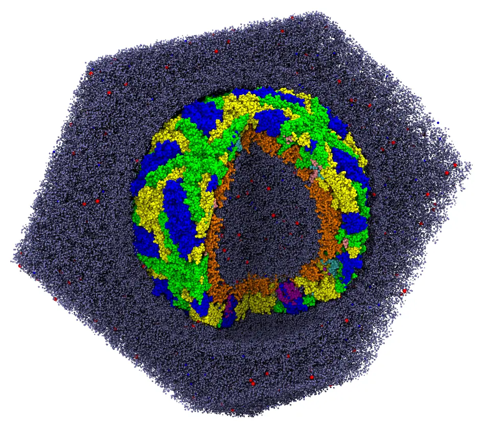
The virion with a quarter cut out: the protein shell (the proteins E in blue, green and yellow, the proteins M in purple, pink and cyan), the two layers of the membrane (orange), water (gray) and ions (red and blue). The water outside is shown only behind the virion.
The files are the packmol_zika set of MolSimTestFiles: download and extract packmol_zika.tar.gz (7 MB), which contains the input file zika.inp and the structures: capsid.pdb (the protein shell, 489960 particles), popc.pdb, pope.pdb and pops.pdb (the lipids), wt4.pdb and wls.pdb (two models of water of SIRAH), and NaW.pdb and ClW.pdb (the ions).
The regions of the system
Everything is organized in spherical shells around the center of the protein shell, which is fixed at the origin. The proteins occupy the region between about 150 and 250 Å from the center, mostly between 177 and 230 Å, with transmembrane segments crossing the membrane:
| Component | Region (distance from the center, Å) |
|---|---|
| Inner layer of the membrane | heads (bead 1) below 160, ends of the chains above 170 |
| Outer layer of the membrane | ends of the chains below 175, heads above 180 |
| Water inside the membrane | WT4 between 120 and 160, WLS below 150 |
| Water between the membrane and the proteins | WT4 between 175 and 195 |
| Water outside | WT4 between 230 and 280, WLS outside 230 |
| Ions | inside 180, between 180 and 215, and outside 220 |
The periodic cell is a rhombic dodecahedron (pbc dodecahedral 0. 0. 0. 674., see Rhombic dodecahedron box): its periodic images are as far apart as those of a cubic box with sides of 674 Å, but it has 71% of the volume of that box, and the water and the ions outside the virion are only those that fill it. The regions outside the virion need no other constraint: the periodic cell bounds them.
The lipids are oriented only by their extreme beads: the head (bead 1) and the end of a chain (bead 16, or 17 in POPS), which are 21 to 25 Å apart. The surfaces that bound them are 10 Å apart in the inner layer and 5 Å apart in the outer one, less than half the length of the lipids, which leaves them room to bend and tilt while being packed.
The virion
The input file, zika.inp:
The input file zika.inp (click to expand)
#
# A Zika virion: the capsid (fixed), surrounded by a membrane of two layers of
# POPC, POPE and POPS, solvated by water and ions inside, between the capsid
# and the membrane, and outside. Coarse-grained (SIRAH) models: WT4 and WLS
# water, NaW and ClW ions.
#
tolerance 2.0
output zika.pdb

# The periodic cell: a rhombic dodecahedron centered at the origin, with 674 Å
# between opposite faces (and between periodic images). The output is written in
# the shape of the dodecahedron.
pbc dodecahedral 0. 0. 0. 674.
periodic_boundary_style dodecahedral

# Uncomment to write only the initial placement of the molecules to the output file,
# without packing them (to check the constraints):
# check_initial_placement

structure capsid.pdb
  number 1
  center
  fixed 0. 0. 0. 0. 0. 0.
end structure

#
# Outer layer of the membrane: heads (bead 1) outside r = 180 Å, ends of the
# chains (bead 16, or 17 in POPS) inside r = 175 Å
#
structure popc.pdb
  number 2185
  atoms 16
    inside sphere 0. 0. 0. 175.
  end atoms
  atoms 1
    outside sphere 0. 0. 0. 180.
  end atoms
end structure

structure pope.pdb
  number 1079
  atoms 16
    inside sphere 0. 0. 0. 175.
  end atoms
  atoms 1
    outside sphere 0. 0. 0. 180.
  end atoms
end structure

structure pops.pdb
  number 367
  atoms 17
    inside sphere 0. 0. 0. 175.
  end atoms
  atoms 1
    outside sphere 0. 0. 0. 180.
  end atoms
end structure

#
# Inner layer of the membrane: heads inside r = 160 Å, ends of the chains
# outside r = 170 Å
#
structure popc.pdb
  number 2855
  atoms 16
    outside sphere 0. 0. 0. 170.
  end atoms
  atoms 1
    inside sphere 0. 0. 0. 160.
  end atoms
end structure

structure pope.pdb
  number 1441
  atoms 16
    outside sphere 0. 0. 0. 170.
  end atoms
  atoms 1
    inside sphere 0. 0. 0. 160.
  end atoms
end structure

structure pops.pdb
  number 473
  atoms 17
    outside sphere 0. 0. 0. 170.
  end atoms
  atoms 1
    inside sphere 0. 0. 0. 160.
  end atoms
end structure

#
# Water inside the membrane: around and within the capsid
#
structure wt4.pdb
  number 17903
  inside sphere 0. 0. 0. 160.
  outside sphere 0. 0. 0. 120.
end structure

structure wls.pdb
  number 23500
  inside sphere 0. 0. 0. 150.
end structure

#
# Water between the two layers of heads and the outer water
#
structure wt4.pdb
  number 7551
  outside sphere 0. 0. 0. 175.
  inside sphere 0. 0. 0. 195.
end structure

structure wt4.pdb
  number 85127
  outside sphere 0. 0. 0. 230.
  inside sphere 0. 0. 0. 280.
end structure

structure wls.pdb
  number 259500
  outside sphere 0. 0. 0. 230.
end structure

#
# Ions: inside, around the membrane, and outside
#
structure NaW.pdb
  number 616
  inside sphere 0. 0. 0. 180.
end structure

structure NaW.pdb
  number 256
  outside sphere 0. 0. 0. 180.
  inside sphere 0. 0. 0. 215.
end structure

structure NaW.pdb
  number 2078
  outside sphere 0. 0. 0. 220.
end structure

structure ClW.pdb
  number 531
  inside sphere 0. 0. 0. 180.
end structure

structure ClW.pdb
  number 167
  outside sphere 0. 0. 0. 180.
  inside sphere 0. 0. 0. 215.
end structure

structure ClW.pdb
  number 1592
  outside sphere 0. 0. 0. 220.
end structure
Check the initial placement first
For a system of this size, checking the initial placement is essential: the packing takes about a minute, but the initial placement takes about 15 seconds (see Checking the initial placement). Uncomment the check_initial_placement line of the input file, and run it:
packmol zika.inpThe initial approximation is written to the output file (zika.pdb). Cut as the image above, it shows that every component is in its region:
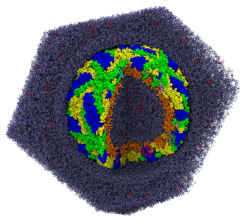
The initial approximation: the molecules are in their regions, but may overlap.
Only then comment the line again, and run the packing.
Packing
packmol zika.inpThe packing converged in 16 to 23 loops, which took about 50 seconds with 14 threads, using 3.5 GB of memory (the check of the initial approximation takes 15 seconds and 3.5 GB). By default, the packmol executable uses all the cores of the machine; packmol -t N limits it to N threads.
- Check the initial placement first (
check_initial_placement): mistakes in the constraints of a system of this size are expensive to find after the packing. restart_fromreads the coordinates of a previous output back, to pack the system in parts: for example, the membrane first, and then the solvent around it (see Restarting a run).- The memory and the time grow roughly linearly with the number of particles.
Julia script
The same system, built in Julia, with the regions of the lipids given once for each layer:
using Packmolcapsid = structure_type("capsid.pdb"; number = 1, fixed = ([0., 0., 0.], [0., 0., 0.]), center = :geometric,)# The lipids of a layer: head (bead 1) and end of the chain# (bead 16, or 17 in POPS)function layer(file, number, tail, head_region, tail_region) structure_type(file; number, atom_constraints = [[1] => head_region, [tail] => tail_region])endorigin = [0., 0., 0.]outer = (OutsideSphere(origin, 180.), InsideSphere(origin, 175.))inner = (InsideSphere(origin, 160.), OutsideSphere(origin, 170.))lipids = [ layer("popc.pdb", 2185, 16, outer...), layer("pope.pdb", 1079, 16, outer...), layer("pops.pdb", 367, 17, outer...), layer("popc.pdb", 2855, 16, inner...), layer("pope.pdb", 1441, 16, inner...), layer("pops.pdb", 473, 17, inner...),]solvent = [ structure_type("wt4.pdb"; number = 17903, constraints = [ InsideSphere(origin, 160.), OutsideSphere(origin, 120.)]), structure_type("wls.pdb"; number = 23500, constraints = [InsideSphere(origin, 150.)]), structure_type("wt4.pdb"; number = 7551, constraints = [ OutsideSphere(origin, 175.), InsideSphere(origin, 195.)]), structure_type("wt4.pdb"; number = 85127, constraints = [ OutsideSphere(origin, 230.), InsideSphere(origin, 280.)]), structure_type("wls.pdb"; number = 259500, constraints = [OutsideSphere(origin, 230.)]),]ions = [ structure_type(ion; number, constraints) for (ion, numbers) in ( ("NaW.pdb", (616, 256, 2078)), ("ClW.pdb", (531, 167, 1592)), ) for (number, constraints) in zip(numbers, ( [InsideSphere(origin, 180.)], [OutsideSphere(origin, 180.), InsideSphere(origin, 215.)], [OutsideSphere(origin, 220.)], ))]system = PackmolSystem([capsid; lipids; solvent; ions]; output = "zika.pdb", tolerance = 2.0, unitcell = dodecahedral_unitcell(Float64, 674.0), # centered at the origin # check_initial_placement = true, # uncomment to only place the molecules)# Write the output in the shape of the rhombic dodecahedrontriclinic_to_dodecahedral(system)packmol(system)